
 Visium
Human liver
GSM9986439
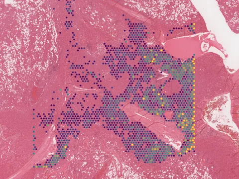
human liver hepatic angiomyolipoma 95 MB 4,988 obs bronze H&E
TechnologyVisium
Organismhuman
Tissueliver
Conditionhepatic angiomyolipoma
Obs4,988
bronze H&E
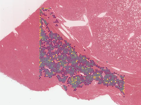
Hepatic angiomyolipoma is a rare mesenchymal tumor frequently associated with tuberous sclerosis complex gene mutations. The tumor microenvironment profiling enables the discovery of potential therapeutic targets. Here, we profile 18,214 cells from tumor and adjacent tissues to identify distinct cell subsets, validate perivascular epithelioid cell biomarkers, predict cellular origins, and characterize molecular networks. Complementarily, we applied microarray-based spatial transcriptomics to map tissue architecture on three hepatic angiomyolipoma specimens by spot arrays. Tumor and normal regions were annotated using robust cell type decomposition and multimodal intersection analysis, integrating snRNA-seq and spatial transcriptomics data. We demonstrate that the perivascular epithelioid cell share substantial transcriptional similarity with endothelial cells and and show a continuous differentiation trajectory. This interaction might be regulated by PDGFD-PDGFRB signaling promotes autophagy by activating the Ras pathway potentially contributing to the pathogenesis and progression of hepatic angiomyolipoma. Our results provide a potential therapeutic strategy by disrupting perivascular epithelioid cell and endothelial cells interaction to improve therapeutic effect.
import lamindb as ln
import spatialdata as sd

coll = ln.Collection.get(key="geo-gse344843")
coll.ordered_artifacts.df()                 # the 3 members, ordered

# download every member .zarr to the local cache:
paths = [a.cache() for a in coll.ordered_artifacts.all()]

# …or load each straight into memory as a SpatialData object:
sdatas = {a.key: sd.read_zarr(a.cache()) for a in coll.ordered_artifacts.all()}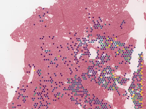 Visium
 Visium
No linked publication found — please cite the data source.