
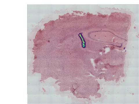
 Visium HD
Mouse brain
GSM9817885
mouse brain 672 MB 384,475 obs bronze H&E
TechnologyVisium HD
Organismmouse
Tissuebrain
Obs384,475
bronze H&E
Spatial transcriptomics on WT versus CADASIL mouse model with brain and isolated vasculature
import lamindb as ln
import spatialdata as sd

coll = ln.Collection.get(key="geo-gse335702")
coll.ordered_artifacts.df()                 # the 8 members, ordered

# download every member .zarr to the local cache:
paths = [a.cache() for a in coll.ordered_artifacts.all()]

# …or load each straight into memory as a SpatialData object:
sdatas = {a.key: sd.read_zarr(a.cache()) for a in coll.ordered_artifacts.all()}Cerebral hemodynamic dysfunction is a key driver of unhealthy brain aging. Impaired microcirculatory reactivity leads to uneven perfusion, rendering deeper brain regions more vulnerable and thereby contributing to cognitive decline. Yet how capillaries contribute to these deficits remains poorly defined. Here we combined spatial transcriptomics with in vivo two-photon and three-photon imaging to measure layer-specific cerebral blood flow in control and small vessel disease model mice (CADASIL TgNotch3R169C). We found downregulation of ATP-synthesizing genes, indicating microvascular metabolic impairment that paralleled impaired pericyte bioenergetics. This energy deficit coincided with diminished tone in the arteriole-capillary transitional zone and reduced deep-layer perfusion. Complementary electrophysiology, ex vivo and in silico approaches, revealed that hyperactive K<sub>ATP</sub> channels in pericytes drive a redistribution of cerebral blood flow toward superficial cortical layers. This loss of spatial perfusion equalization, despite preserved global flow, contributed to deep-layer hypoperfusion, establishing a previously underrecognized but tractable vascular function disrupted in aging pathology.
Visium HD 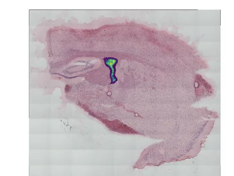 Visium HD
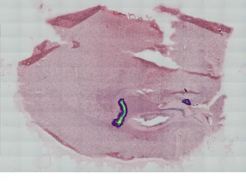 Visium HD
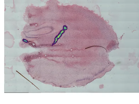 Visium HD
 Visium
 Visium
 Visium
 Visium
@article{Jeffrey_2026,
  title = {Pericyte KATP channel hyperactivity redistributes cortical blood flow in a CADASIL mouse model},
  volume = {5},
  ISSN = {2731-0590},
  url = {http://dx.doi.org/10.1038/s44161-026-00844-0},
  DOI = {10.1038/s44161-026-00844-0},
  number = {8},
  journal = {Nature Cardiovascular Research},
  publisher = {Springer Science and Business Media LLC},
  author = {Jeffrey, Danielle A. and Prince, Eric W. and Khakpour, Niloufar and Ferris, Hannah R. and Peters, Colin H. and Seedorf, Gregory and Romero, Phinea Z. and Bueno Guerrero, Mayra and Russell, Abigail N. and Glodoski, Katherine and Futia, Gregory L. and Bonney, Stephanie K. and Thornton, Michael A. and Gibson, Emily A. and Proenza, Catherine and Garcia, Anastacia M. and Tsoukias, Nikolaos M. and Dabertrand, Fabrice},
  year = {2026},
  month = Aug,
  pages = {725–743}
}