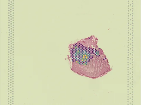
 Visium
Human skin of body
GSM9483975
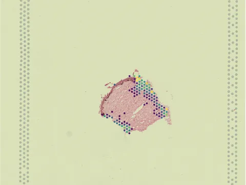
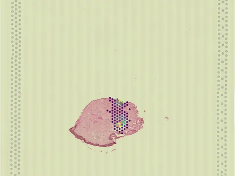
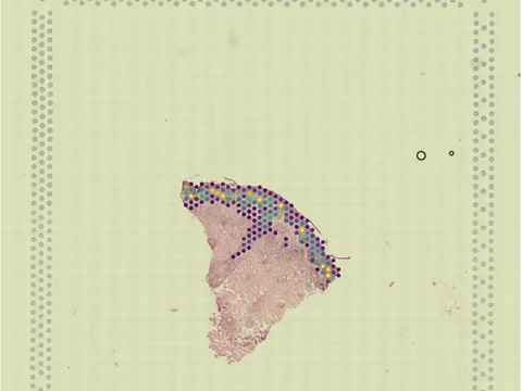
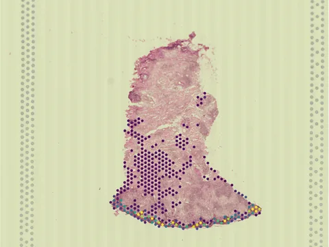
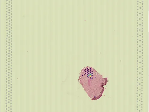
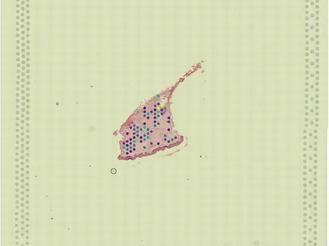
human skin of body cutaneous leishmaniasis 11 MB 544 obs bronze H&E
TechnologyVisium
Organismhuman
Tissueskin of body
Conditioncutaneous leishmaniasis
Obs544
bronze H&E
Ethiopian cutaneous leishmaniasis (CL) shows remarkably heterogeneous clinical presentations, but the underlying immunopathological mechanisms driving this heterogeneity in disease presentation remains poorly understood. To characterise the local immune response in Ethiopian CL, we performed spatial transcriptomics (Visium, 10x Genomics) on paired (lesional and non lesional) skin punch biopsies from five Ethiopian CL patients. We used reference-free deconvolution, morphology-guided regional analyses, and immunohistochemistry to identify five5 dominant patient-defined immunopathological responses: i) epithelial hyperplasia with interferon-stimulated keratinocytes, ii) cytotoxicity with tertiary lymphoid structures, iii) granulomatous inflammation with proinflammatory response iv) granulomatous inflammation with M2-polarised myeloid cell responses , and v) fibrotic remodelling with active collagen synthesis. Future longitudinal studies are needed to distinguish whether these patterns represent discrete pathotypes or stages of disease progression. Improved understanding of disease pathotypes in Ethiopian CL directly supports the WHO 2030 roadmap for leishmaniasis control and informs future immune-based therapeutic strategies.
import lamindb as ln
import spatialdata as sd

coll = ln.Collection.get(key="geo-gse317987")
coll.ordered_artifacts.df()                 # the 10 members, ordered

# download every member .zarr to the local cache:
paths = [a.cache() for a in coll.ordered_artifacts.all()]

# …or load each straight into memory as a SpatialData object:
sdatas = {a.key: sd.read_zarr(a.cache()) for a in coll.ordered_artifacts.all()}
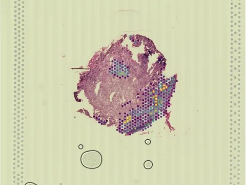 Visium
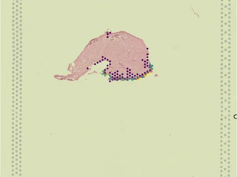 Visium
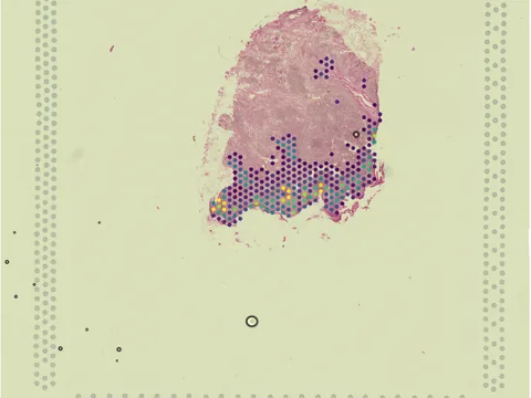 Visium
 Visium
 Visium
 Visium
 Visium
 Visium
 Visium
No linked publication found — please cite the data source.