
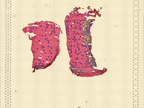
 Visium
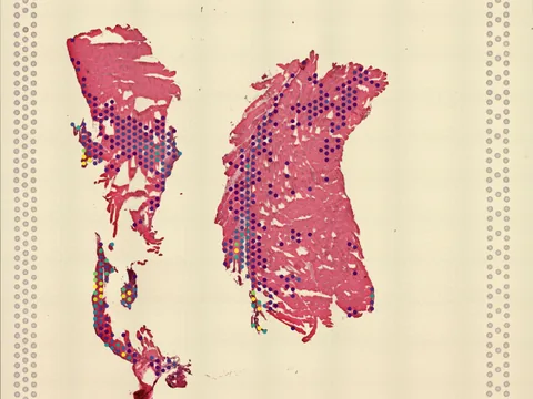
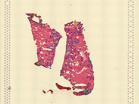
Mouse plantaris
GSM8919244
mouse plantaris 14 MB 1,068 obs bronze H&E
TechnologyVisium
Organismmouse
Tissueplantaris
Obs1,068
Magnificationother · 3.497 µm/px
bronze H&E
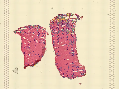
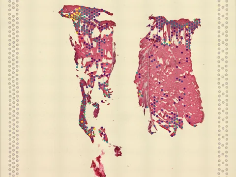
The molecular mechanisms underlying the opposing states of muscle atrophy (such as with disuse or in aging) and hypertrophy (such as with increased physical activity) are still not well-defined. Progress has been slowed by challenges in characterizing the niche heterogeneity of skeletal muscle (whereby myofibers are the most abundant) and in obtaining well-characterized samples. Here we describe: i) changes in muscle cell populations that are undergoing atrophy or hypertrophy, with complex, opposing cell transcriptional responses during each process; and ii) a remarkable remodeling of the neuromuscular junction (NMJ) domain, whereby we identify emerging new subpopulations of synaptic myonuclei (SynM) and terminal Schwann cells (tSCs) that are specifically adapted to each growth/atrophy condition. High-resolution 3D imaging and spatial transcriptomics reveal these structural and molecular adaptations at the tissue level. Critically, these changes also occur in the NMJ of both denervated and exercised human muscle. Our study lays the ground for identifying novel targets for medical, pharmacological and lifestyle interventions in aging and disease.
import lamindb as ln
import spatialdata as sd

coll = ln.Collection.get(key="geo-gse294765")
coll.ordered_artifacts.df()                 # the 6 members, ordered

# download every member .zarr to the local cache:
paths = [a.cache() for a in coll.ordered_artifacts.all()]

# …or load each straight into memory as a SpatialData object:
sdatas = {a.key: sd.read_zarr(a.cache()) for a in coll.ordered_artifacts.all()}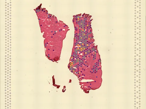 Visium
 Visium
 Visium
 Visium
 Visium
No linked publication found — please cite the data source.