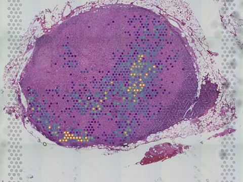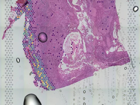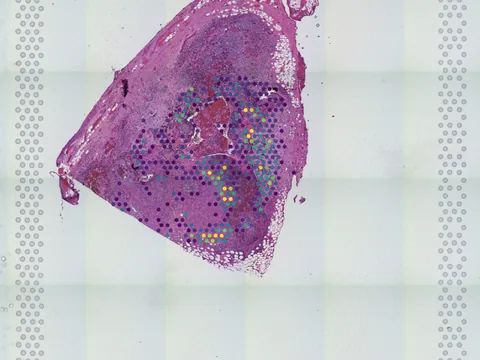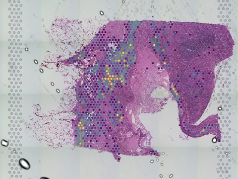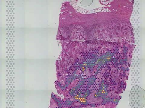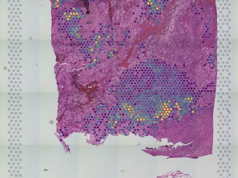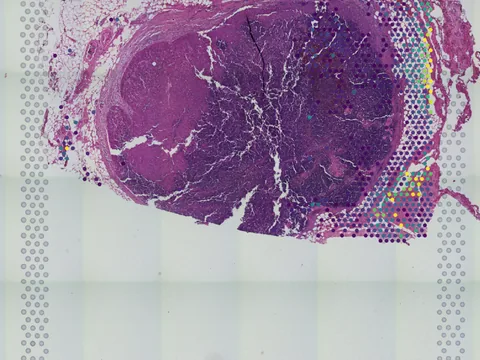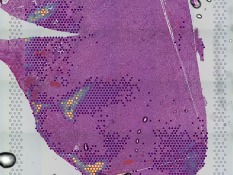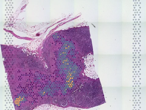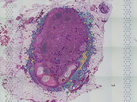
Collections / Cancer-induced nerve injury promotes resistance to anti-PD-1 therapy [Spatial Transcriptomics]

Cancer-induced nerve injury promotes resistance to anti-PD-1 therapy [Spatial Transcriptomics]

Peri-neural invasion (PNI) is a well-established poor prognostic factor in multiple cancer types. However, the mechanisms driving the PNI's detrimental clinical effect remain elusive. Here, we provide clinical and mechanistic insights into PNI and cancer-induced injury of tumor-associated nerves (TANs) and their role in resistance to anti-PD-1 therapy. Our work demonstrates that poor response to anti-PD-1 therapy in cutaneous squamous cell carcinoma (cSCC), melanoma, and gastric cancer is associated with PNI and TANs injury. Ultrastructural electron microscopy analysis reveals that direct contact between cancer cells and nerve fibers leads to cancer-induced nerve injury (CINI) via myelin degradation. Injured neurons respond by autonomously initiating an interleukin (IL)-6 and interferon (IFN) type I inflammatory response. This inflammatory response alters the immune activity in the peri-neural niche in melanoma, cSCC, and pancreatic adenocarcinoma, leading to an immuno-suppressive activity aimed at nerve healing and regeneration. As the tumor grows, the CINI burden increases, the inflammatory signal within the niche becomes chronic, and eventually skews the general immune tone within the tumor microenvironment to a suppressive and exhaustive state. The CINI-driven anti-PD-1 resistance can be reversed by targeting multiple steps in the CINI signaling process: denervating the tumor, conditional knockout of the transcription factor mediating the injury signal within neurons (cKO-Atf3), knockout of the IFN-a receptor signaling (Ifnar1-/-), or by combining anti-PD-1 and anti-IL-6-receptor blockade. Our findings demonstrate the direct immuno-regulatory roles of TANs and their therapeutic potential.

Datasets11
Size~448 MB
AssayVisium
Samplehuman · skin of body
KindOther
PMID:40836096 ↗
Python · lamindb
import lamindb as ln
import spatialdata as sd

coll = ln.Collection.get(key="geo-gse289745")
coll.ordered_artifacts.df()                 # the 11 members, ordered

# download every member .zarr to the local cache:
paths = [a.cache() for a in coll.ordered_artifacts.all()]

# …or load each straight into memory as a SpatialData object:
sdatas = {a.key: sd.read_zarr(a.cache()) for a in coll.ordered_artifacts.all()}
Open in Vitessce
Visium
Human skin of body
GSM8797982
human skin of body skin squamous cell carcinoma 45 MB 2,163 obs bronze H&E
TechnologyVisium
Organismhuman
Tissueskin of body
Conditionskin squamous cell carcinoma
Size45 MB
Obs2,163
Magnificationother · 3.529 µm/px
bronze H&E Open in Vitessce
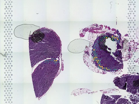
Open in Vitessce
Visium
Human skin of body
GSM8797974
human skin of body skin squamous cell carcinoma 28 MB 1,274 obs bronze H&E
TechnologyVisium
Organismhuman
Tissueskin of body
Conditionskin squamous cell carcinoma
Size28 MB
Obs1,274
Magnificationother · 3.433 µm/px
bronze H&E Open in Vitessce
Open in Vitessce
Visium
Human skin of body
GSM8797979
human skin of body skin squamous cell carcinoma 37 MB 2,779 obs bronze H&E
TechnologyVisium
Organismhuman
Tissueskin of body
Conditionskin squamous cell carcinoma
Size37 MB
Obs2,779
Magnificationother · 3.446 µm/px
bronze H&E Open in Vitessce
Open in Vitessce
Visium
Human skin of body
GSM8797973
human skin of body skin squamous cell carcinoma 33 MB 2,895 obs bronze H&E
TechnologyVisium
Organismhuman
Tissueskin of body
Conditionskin squamous cell carcinoma
Size33 MB
Obs2,895
Magnificationother · 3.506 µm/px
bronze H&E Open in Vitessce
Open in Vitessce
Visium
Human skin of body
GSM8797975
human skin of body skin squamous cell carcinoma 17 MB 1,514 obs bronze H&E
TechnologyVisium
Organismhuman
Tissueskin of body
Conditionskin squamous cell carcinoma
Size17 MB
Obs1,514
Magnificationother · 3.476 µm/px
bronze H&E Open in Vitessce
Open in Vitessce
Visium
Human skin of body
GSM8797980
human skin of body skin squamous cell carcinoma 26 MB 2,063 obs bronze H&E
TechnologyVisium
Organismhuman
Tissueskin of body
Conditionskin squamous cell carcinoma
Size26 MB
Obs2,063
Magnificationother · 3.478 µm/px
bronze H&E Open in Vitessce
Open in Vitessce
Visium
Human skin of body
GSM8797981
human skin of body skin squamous cell carcinoma 59 MB 2,748 obs bronze H&E
TechnologyVisium
Organismhuman
Tissueskin of body
Conditionskin squamous cell carcinoma
Size59 MB
Obs2,748
bronze H&E Open in Vitessce
Open in Vitessce
Visium
Human skin of body
GSM8797978
human skin of body skin squamous cell carcinoma 56 MB 3,195 obs bronze H&E
TechnologyVisium
Organismhuman
Tissueskin of body
Conditionskin squamous cell carcinoma
Size56 MB
Obs3,195
Magnificationother · 3.503 µm/px
bronze H&E Open in Vitessce
Open in Vitessce
Visium
Human skin of body
GSM8797977
human skin of body skin squamous cell carcinoma 54 MB 2,887 obs bronze H&E
TechnologyVisium
Organismhuman
Tissueskin of body
Conditionskin squamous cell carcinoma
Size54 MB
Obs2,887
Magnificationother · 3.525 µm/px
bronze H&E Open in Vitessce
Open in Vitessce
Visium
Human skin of body
GSM8797983
human skin of body skin squamous cell carcinoma 61 MB 4,271 obs bronze H&E
TechnologyVisium
Organismhuman
Tissueskin of body
Conditionskin squamous cell carcinoma
Size61 MB
Obs4,271
bronze H&E Open in Vitessce
Open in Vitessce
Visium
Human skin of body
GSM8797976
human skin of body skin squamous cell carcinoma 32 MB 2,041 obs bronze H&E
TechnologyVisium
Organismhuman
Tissueskin of body
Conditionskin squamous cell carcinoma
Size32 MB
Obs2,041
bronze H&E Open in Vitessce

Cite this collection

No linked publication found — please cite the data source.