
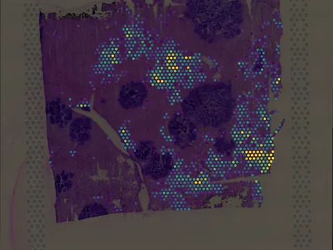
 Visium
Mouse liver
GSM6661400
mouse liver 29 MB 3,751 obs bronze H&E
TechnologyVisium
Organismmouse
Tissueliver
Obs3,751
Magnificationother · 4.176 µm/px
bronze H&E
The spatial organization of cells within tissues is tightly linked to their biological function. Yet, methods to probe the entire transcriptome of multiple native tissue microenvironments at single cell resolution are lacking. Here, we introduce fragment-sequencing, a method that enables the transcriptomic characterization of single cells within spatially distinct tissue niches. Fragment-sequencing of the mouse metastatic liver revealed previously uncharacterized zonated genes and ligand-receptor interactions enriched in the different hepatic microenvironments and the metastatic niche.
import lamindb as ln
import spatialdata as sd

coll = ln.Collection.get(key="geo-gse216189")
coll.ordered_artifacts.df()                 # the 2 members, ordered

# download every member .zarr to the local cache:
paths = [a.cache() for a in coll.ordered_artifacts.all()]

# …or load each straight into memory as a SpatialData object:
sdatas = {a.key: sd.read_zarr(a.cache()) for a in coll.ordered_artifacts.all()}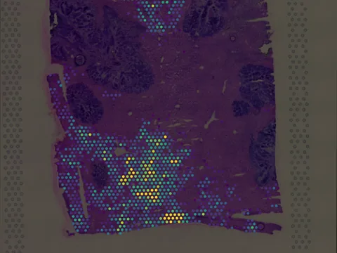 Visium
No linked publication found — please cite the data source.